Student in the Lab : Guts and Glory
Photography by Chantae Bennett, undergraduate student,
Harriet L. Wilkes Honors College
Harriet L. Wilkes Honors College
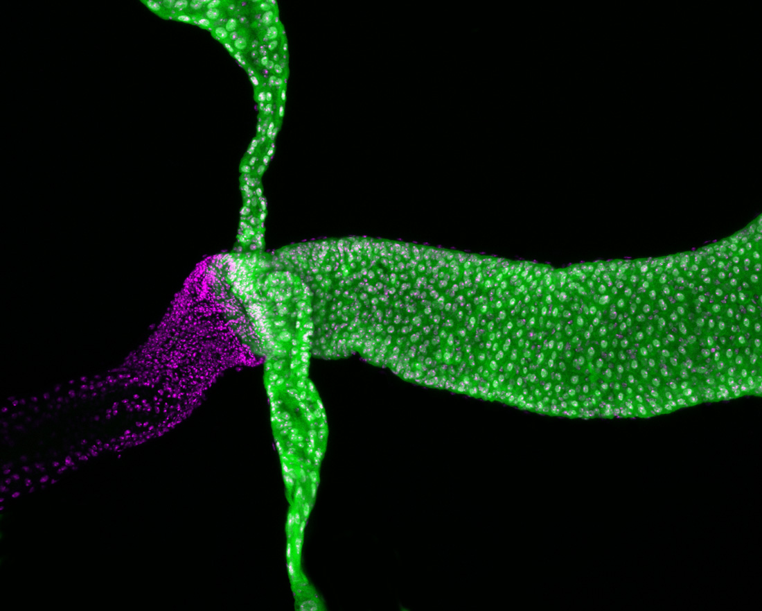
This image shows the renal tubes, the hindgut, and the midgut of fruit fly. Nuclei of all cells were labeled with DAPI dye (magenta). Antibody labeling against the human Attractin homolog (green) shows that the protein is expressed in the renal tubes and midgut but not in the hindgut. Image was taken with a 20x objective using a Nikon AR1 confocal microscope.