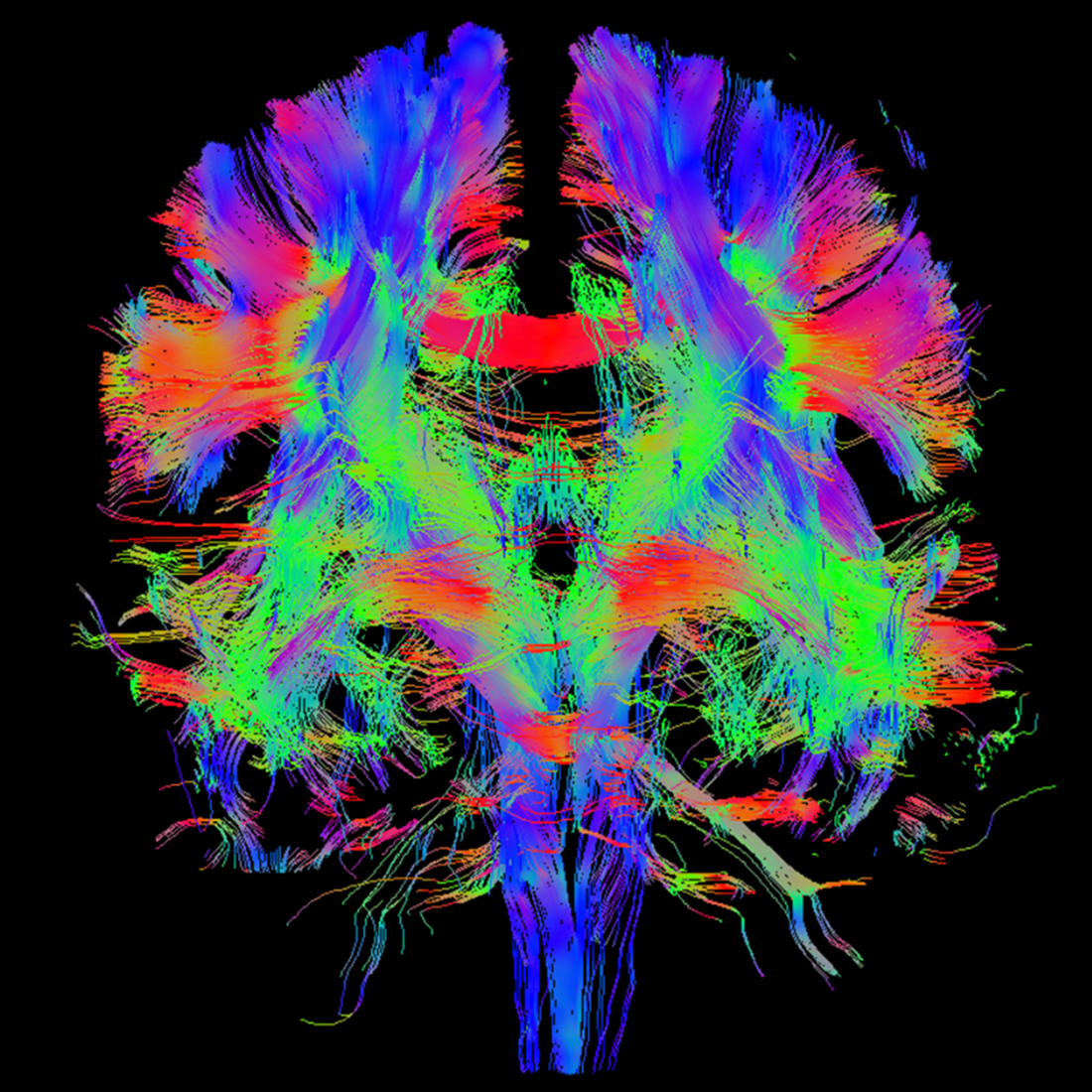
Honorable Mention: Brain Fibers
Division of Research
White matter fiber tracts in the brain, identified using diffusion MRI, reveal the pathways of microscopic water movement. The fibers are color-coded according to the direction of water diffusion: Blue represents diffusion in the up-down (inferior-superior) direction, green indicates diffusion in the front-back (anterior-posterior) direction, and red shows diffusion in the left-right direction. The MRI data were acquired from a healthy volunteer at Florida Atlantic University's MRI facility. Magnetic resonance imaging (MRI) is a non-ionizing medical imaging technique that uses magnetic fields to generate detailed images. In this case, MRI was employed to capture a series of brain images, each sensitive to water movement along a specific direction. Mathematical models were then used to compute the direction and magnitude of the brain fibers. A tracing technique was applied to identify the white matter fiber tracts, with the visualization color-coding the fibers based on the direction of diffusion.